
Mice are used a lot in scientific research, usually in lab tests. Now, researchers in Germany have made it possible to look at a mouse’s insides by making the animals transparent.
Ali Ertürk, a brain researcher at Ludwig Maximilians University of Munich and a team, have used a special method that removes liquids and fats from tissue, making mice “see-through,” NBC News reports.
The researchers hope this revolutionary method can be used to make whole, unsliced models of the human brain, with all of the nerves untouched. They also say this development will allow scientists around the world to study mice without having to kill them.
Ertürk says,
Now, for the first time, we have a powerful tool that can make the human brain transparent and reduce its size to fit an imaging microscope for mapping.
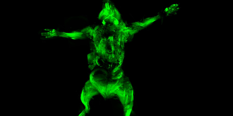
The process is called ultimate DISCO, short for 3D imaging of solvent-cleared organs. It does two things: makes it possible to see whole systems intact, and shrinks the mouse so that it fits under a microscope.
The study expects that this method can also be applied to small monkeys, and to whole brains in the future. “We all know the big fuss (rightfully) around mapping the human brain. But so far there is not any approach that even comes close to mapping any part of the human brain at individual neuron level,” Ertürk says.
He adds that this technique will save some lab animals, as research usually has to sacrifice a whole animal for small tissue samples.
Ertürk believes that using uDISCO, “scientists can start to build whole body atlases for various biological systems such nerves, vasculature and immune cells innervating whole body. This will at least provide the basic knowledge about how a healthy organ is organized in terms of these mapped systems (nerves, vessels, etc.).” He says all of this valuable knowledge can then be available to everyone.
To get their procedure right, the researchers anesthetized live mice. They used several agents to make it possible to see the systems in images when fluorescent light illuminated them, or by other processes — similar to how a contrast dye is used for an MRI.
Once the dye was pumped through the mice’s tissue, they were killed and their tissues cleared of water and fat. Water and fat combined scatter light, making the tissue stand out and clearly visible.
Scientists have long been using very thin slices of organs in imaging and recreating 3D computer models. But even the thinnest slices can interrupt neurons and other connections, which might compromise important information. This way, any organs that undergo the process can be studied whole, which is important in studying challenging diseases like Alzheimer’s or mental illnesses.
The method was published in the journal Nature Methods.